
Hope Is Here
For Doctors & Cancer Patients
Cancer patients and their doctors often face agonizing trade-offs in the fight against cancer ‒ but hope is here. Cutting edge technology from Proton Calibration Technologies (PCT) puts more options at the fingertips of radiation oncologists to help improve patient outcomes while reducing treatment costs. PCT is on a mission to take the fight against cancer to the next level with our precise, proven, and proprietary technology.
Proton computed tomography (pCT) is not only transformative enabling technology for cancer treatment. It will revolutionize how cardiac ablation is performed using proton beams. No longer will patients have to endure hours under anesthesia and internal bleeding. With pCT, durable ablation of cardiac arrhythmia can be performed without anesthesia and in less than half an hour.
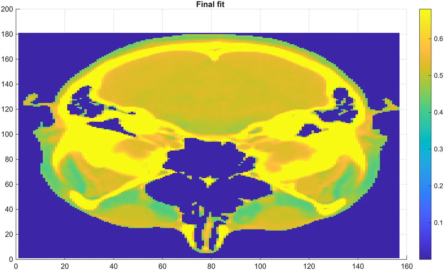
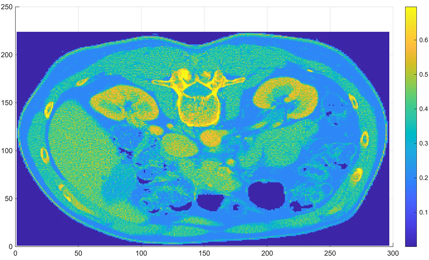
Proton CT Images Created Using Proton Calibration Technologies’ Novel Algorithm
To see how 3D images are created from 2D slices click on the above video.
Pictured above, the algorithm was used to create axial slice proton Computed Tomography (pCT) images of a human head (left) and abdomen (right). The colors represent the proton Relative Stopping Power (RSP) of the various tissues as high-energy protons pass through. Proton energies of 250 MeV were used for the head image, and 330 MeV proton energies were used for the image of the abdomen*. The RSP for each image voxel, one cubic millimeter in size, is calculated by the pCT algorithm from proton energy losses. The Geant4 high energy physics simulation platform was used to determine the energy absorbed by each “ideal proton” (see “How the Algorithm Creates pCT Images” in the News section); the aggregate simulations are then used by the pCT algorithm to create the image – essentially a “RSP map”. This technique is analogous to X-ray CT imaging which measures the radiodensity of tissue in each voxel. Similarly, both methods create the 3D image volume by stacking 2D axial slices.
* Higher energy is required for protons to pass completely through the abdomen. 330 MeV is the highest therapeutic proton energy available from commercial proton therapy systems in use. In the abdominal case the RSP were adjusted to simulate 200 MeV proton energy. The reason for the adjustment is discussed in “How the Algorithm Creates pCT Images” in the News section). The treatment planning system will use the NIST stopping power tables for human tissue to compute the RSP of each voxel as seen by a proton, which continues to lose energy as it travels towards the tumor.
Join Us
In the Fight for faster & safer treatments
Technology
Our key insight is that pCT requires a different imaging algorithm. PCT’s novel, patent-pending proprietary algorithm provides a highly accurate direct measurement of proton stopping powers within 0.1% of the true value for each 1 millimeter voxel within the 3D region of interest. PCT is currently vetting strategic technology partnership opportunities to help make this cutting edge medical imaging modality available to proton beam therapy centers around the world.


Investors
Here is a chance to be in on the next 3D medical imaging system. Looking to the past, we can see that those who recognized the future potential of X-ray CT and MRI early in development were rewarded. Rarely do such ground-floor opportunities occur again. pCT will enable a major advance in the fight against cancer and facilitate the introduction of a new modality to ablate cardiac arrhythmia.
“Proton therapy installations will be keen on acquiring pCT imaging systems as these will significantly increase the precision of treatment, throughput, and therefore overall profitability.”

“RDI will support commercialization of this important pCT technology by developing beam position and fluence imaging instrumentation based on our hardware and reconstruction techniques. ”

“Clinical utility of a pCT system would be in several proton therapy use cases including standard fractionation, hypo-fractionation, stereotactic body RT (SBRT) and stereotactic radiosurgery (SRS).”

“I believe that proton beam is going to play an important role in the ablation of cardiac arrhythmia. In this application, it will be important to eliminate range uncertainty for ablation of a small target volume, and I believe that proton CT will be the key technology needed for imaging and planning this treatment.”